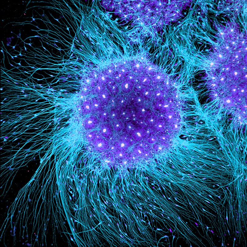
About Meiosis II
In some species, cells enter a brief interphase, or interkinesis, before entering meiosis II. Interkinesis lacks an S phase, so chromosomes are not duplicated. The two cells produced in meiosis I go through the events of meiosis II in synchrony. During meiosis II, the sister chromatids within the two daughter cells separate, forming four new haploid gametes. The mechanics of meiosis II is similar to mitosis, except that each dividing cell has only one set of homologous chromosomes. Therefore, each cell has half the number of sister chromatids to separate out as a diploid cell undergoing mitosis.
Meiosis II Stages
Prophase II

Prophase II is the phase that follows after meiosis I, or after interkinesis if present. If interkinesis takes place, the nuclear envelope and the nucleolus disintegrate during prophase II. The chromosomes are condensed. The centrosomes replicate and move towards the opposite poles. Spindle fibers grow outward from the centrosomes. Prophase II ends where metaphase II begins.
The difference between prophase I and prophase II is that crossing over between chromosomes takes place only in prophase I, not on prophase II.
Source: https://www.biologyonline.com/dictionary/prophase-ii
Metaphase II
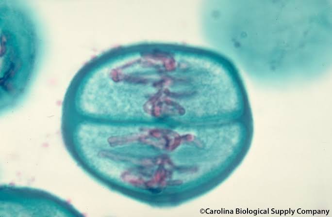
Metaphase II is the second stage in meiosis II. It follows prophase II, which primarily highlights the condensation of the chromosomes and the movement of centrosomes to polar regions of the cell. The cell is in metaphase II when the chromosomes align themselves along the metaphase plate through the facilitation of the spindle fibers. The spindle fibers are now attached to the two kinetochores contained in the centromere of each chromosome. Similar to mitotic metaphase, the two kinetochores are bound to the spindle fibers rom opposite poles and they lie on the equatorial plane, readying for the chromosomal movement towards opposite poles in anaphase II.
Source: https://www.biologyonline.com/dictionary/metaphase-ii
Anaphase II
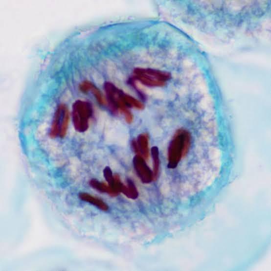
Anaphase II is the third stage in meiosis II. It is the stage after metaphase II, which is that phase wherein the chromosomes are at the equatorial plane and spindle fibers are attached to the kinetochores. Anaphase II is the stage when sister chromatids of every chromosome separate and begin to move towards the opposite ends of the cell. The separation and the movement is due to the shortening of the kinetochore microtubules. Anaphase II precedes telophase II.
Meiotic anaphase II is similar to the anaphase in mitosis. Both mitotic anaphase and meiotic anaphase II involves the separation of sister chromatids towards the opposite poles of the cell. In anaphase I, the paired homologous chromosomes are the ones separating from each other; as a result, the sister chromatids remain together.
Telophase II
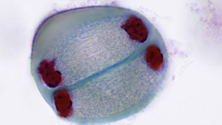
Telophase II is the stage in meiosis II that follows after anaphase II. In anaphase II, the sister chromatids that were formerly joined at the centromere are separated from each other and moved away to opposite poles. At this point, the sister chromatids are sometimes referred to as sister chromosomes. The complete movement and separation of sister chromosomes mark the telophase II. This will then be followed by cytokinesis, wherein each of the two cells produced from meiosis I will give rise to two daughter cells, resulting in a total of four genetically dissimilar haploid cells. The chromosomes de-condense and lengthen. The spindle disassemble and nuclear envelopes reform.
Meiosis II Video Explanation
History of Meiosis
Oscar Hertwig
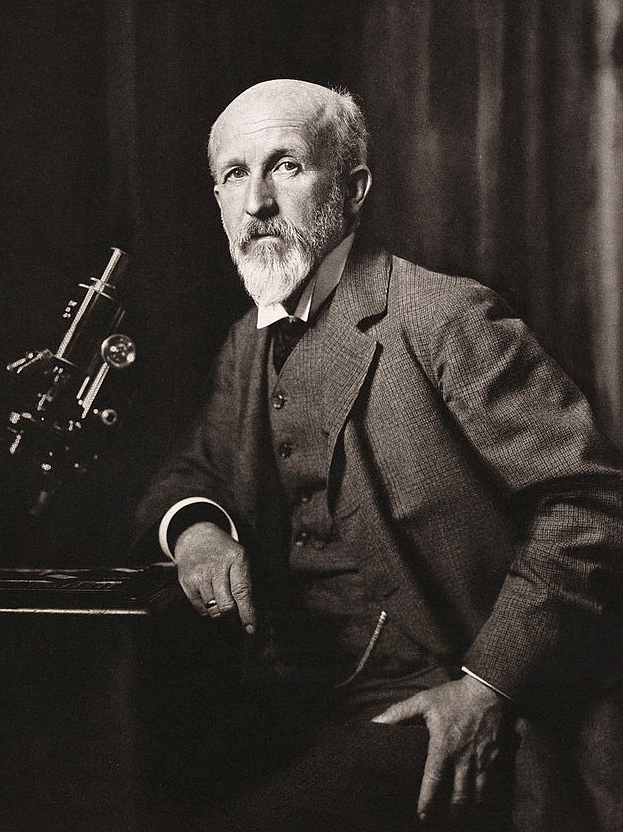
Meiosis was discovered and described for the first time in sea urchin eggs in 1876 by the German biologist Oscar Hertwig. It was described again in 1883, at the level of chromosomes, by the Belgian zoologist Edouard Van Beneden, in Ascaris roundworm eggs.
Source: https://en.m.wikipedia.org/wiki/Meiosis
August Weismann
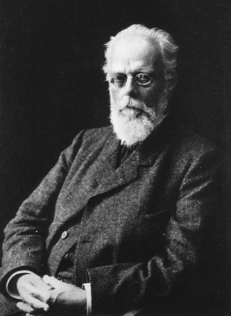
The significance of meiosis for reproduction and inheritance, however, was described only in 1890 by German biologist August Weismann, who noted that two cell divisions were necessary to transform one diploid cell into four haploid cells if the number of chromosomes had to be maintained.
Source: https://en.m.wikipedia.org/wiki/Meiosis
Thomas Hunt Morgan
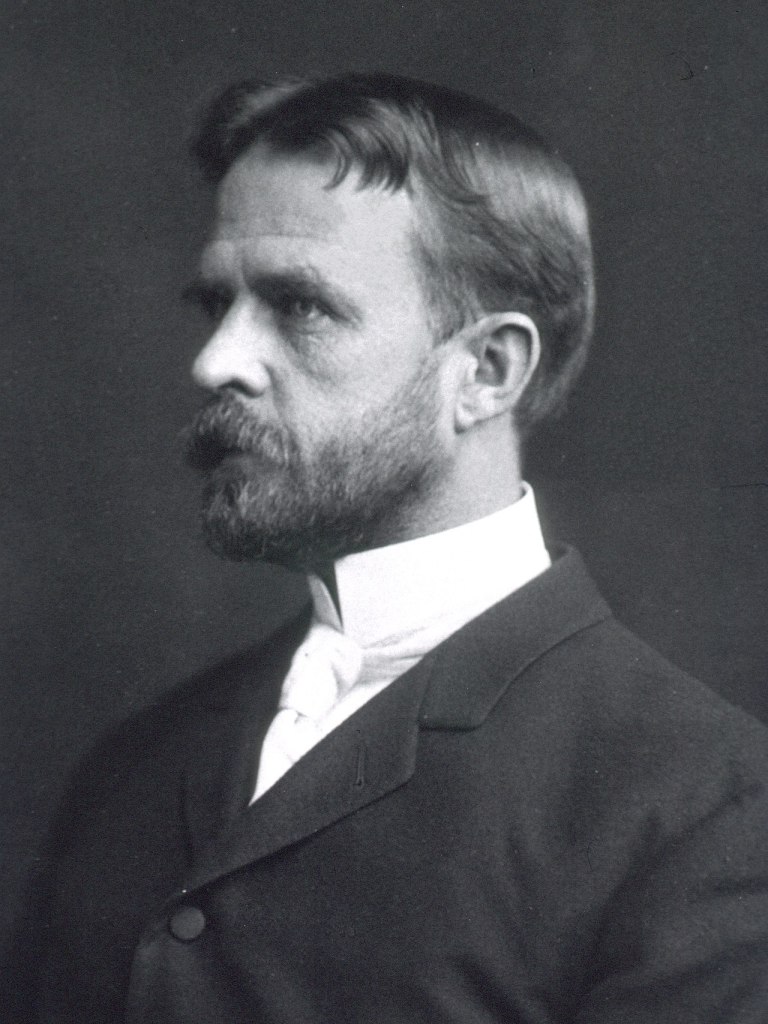
In 1911, the American geneticist Thomas Hunt Morgan detected crossovers in meiosis in the fruit fly Drosophila melanogaster, which helped to establish that genetic traits are transmitted on chromosomes.
Origin
The term “meiosis” is derived from the Greek word μείωσις, meaning ‘lessening’. It was introduced to biology by J.B. Farmer and J.E.S. Moore in 1905, using the idiosyncratic rendering “maiosis”:
Mark Daniel L. Pesigan
11-Commitment
